IVP
Uses
- Your healthcare provider will explain the procedure to you. Ask him or her any questions you have about the procedure.
- You may be asked to sign a consent form that gives permission to do the procedure. Read the form carefully and ask questions if anything is not clear.
- Follow any directions you are given for not eating or drinking before surgery.
- Healthcare provider if you are pregnant or think you may be.
- Healthcare provider if you are allergic to contrast dye or iodine.
- Healthcare provider if you are sensitive to or are allergic to any medicines, latex, tape, or anesthetic medicines (local and general).
- Healthcare provider about all medicines you are taking. This includes prescriptions, over-the-counter medicines, and herbal supplements.
- Healthcare provider if you have had a bleeding disorder. Also tell your provider if you are taking blood-thinning medicine (anticoagulant), aspirin, or other medicines that affect blood clotting.
- You need to take a laxative the night before the test and have a cleansing enema or suppository a few hours before the test.
- You may need to have a blood test to see how well your kidneys will react to the contrast dye.
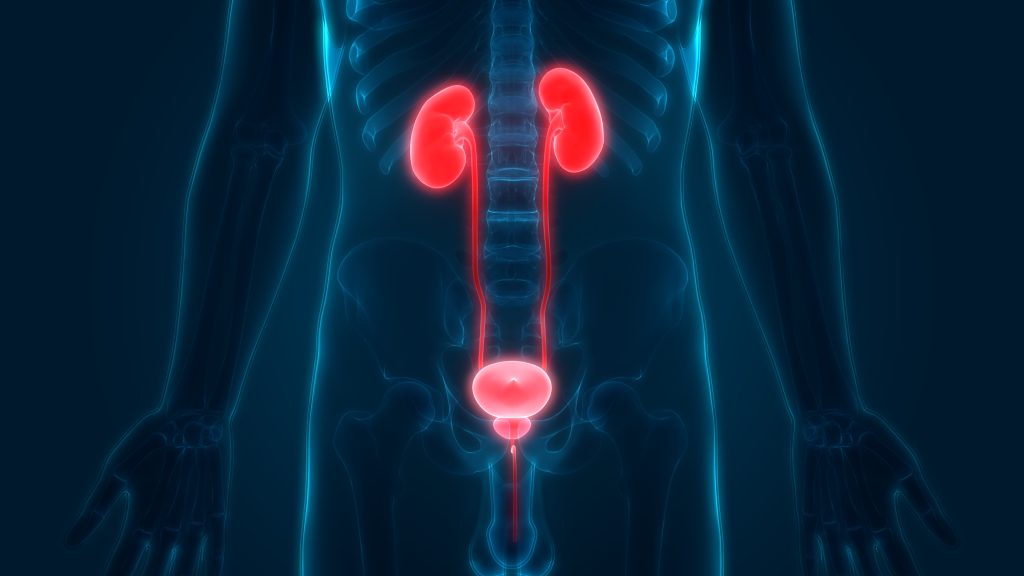
An IVP is an imaging test used to look at the kidneys and ureters. The ureters are the narrow tubes that carry urine from the kidneys to the bladder. During the test, the radiologist injects a contrast dye into one of your veins. He or she uses X-ray images to watch the contrast dye as it moves from the kidney into the ureter and then to the bladder.
Dye that moves too slowly or not at all may mean that you have a blockage in the blood flow through a kidney. It may also mean that the kidney, ureter, or bladder is not working as well as it should. This test may be ordered if your healthcare provider thinks you may have kidney disease or a urinary tract problem.
X-rays use a small amount of radiation to create images of your bones and internal organs. An IVP is one type of X-ray. This test may be done at the same time as a CT scan of the kidneys (urography). The CT scan also uses contrast dye. It makes images that show layers or “slices” of the kidney.

