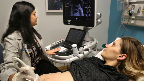
Fetal Echocardiography
Uses
- Assess Heart Structure: Evaluates the heart's anatomy and function to detect congenital heart defects.
- Monitor Heart Function: Checks for any abnormalities in heart rhythm or function.
- Typically Performed: Usually between 18 and 24 weeks of pregnancy, but can be done earlier or later if needed.
- Ultrasound: Uses high-frequency sound waves to create detailed images of the fetal heart.
- Specialized Views: Focuses on obtaining various views of the heart chambers, valves, and blood flow.
- Non-Invasive: Safe for both the mother and the fetus, with no known risks.
Fetal Echocardiography is a specialized ultrasound technique used to examine the heart of a fetus during pregnancy.

